Вирусы
Научная деятельность группы Структурной биотехнологии в области вирусологии ориентирована как на бактериофаги, так и на вирусы эукариот. Исследование вирионов с помощью Крио-ЭМ является сильным дополнением для современных методов анализа геномов. Данные о геометрии и белковом составе необходимы как для возможного биотехнологического применения вирусов, так и потенциального лечения ныне неизлечимых болезней.
Публикации группы по направлению
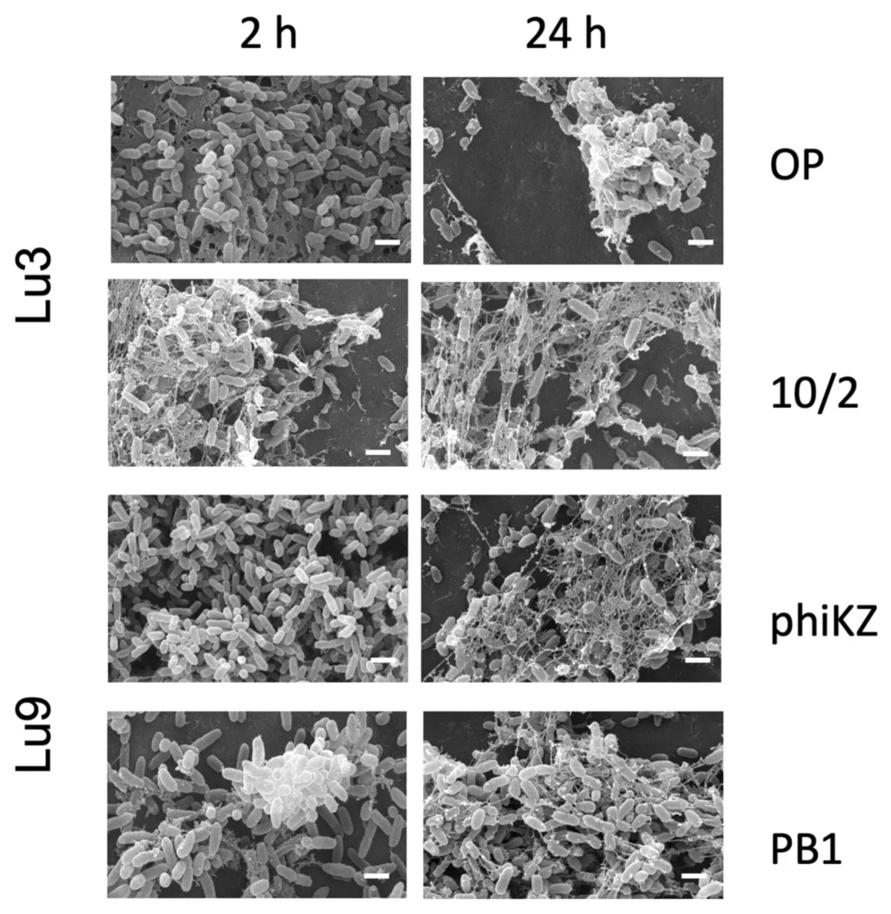
The Efficacy of Giant Phikz Viruses in Degrading Biofilms of Pseudomonas aeruginosa in Comparison to Pbuna Viruses
Wang Y, Bourkaltseva M, Burykin A, Krylov S, Pleteneva E, Krylov V, Sokolova OS
Pseudomonas aeruginosa poses major public health threats, due to its robust, treatment-resistant biofilms, which contribute to multi-drug resistance. Bacteriophages offer a promising alternative. This study evaluates giant Phikzvirus and conventional Pbunavirus phages against pre-formed biofilms from four multi-drug-resistant P. aeruginosa clinical isolates. We tested six phages (three Phikzvirus, three Pbunavirus) against four clinical strains, isolated from chronic urological and pulmonary infections (Ur1, Ur14, Lu3, Lu9) at MOIs 0.001–0.1, quantifying biofilm biomass by crystal violet and visualizing architecture by SEM. Phage treatment leads to significant disrupted biofilms in three isolates achieving more than 50% reduction—comparable with typical antibiotic efficacy against mature biofilms. All biofilms retained their EPS architecture after the treatment, as revealed by SEM, suggesting that residual eDNA-polysaccharide complexes could maintain structural cohesion even after bacterial lysis. The phage’s ability to reduce biofilm biomass demonstrated a significant dependence on the multiplicity of infection (MOI). The best results of anti-biofilm activity of phages (reduction in biofilm biomass by more than 75%) were observed for the phage phiKZ for two strains—Ur1 at MOI = 0.001 and Lu9 at MOI = 0.01—and for the phage phi14/1 for the Ur1 strain at MOI = 0.001. The biofilm formed by the antibiotic-resistant clinical isolate Ur14 demonstrated exceptional resistance to both types of bacteriophages despite the sensitivity of bacteria of this strain to the studied phages. This highlights the need for personalized phage therapy, where tailored phage cocktails are selected for each specific bacterial strain to achieve optimal destruction of the biofilm. DOI
Screening of the G7C Bacteriophage Early Proteins for Antibacterial Activity
S. Y. Rassadina, V. A. Kasyanov, M. Dmitrieva, I. V. Kurdyumova, D. A. Antonova, O. S. Sokolova & M. V. Yakunina
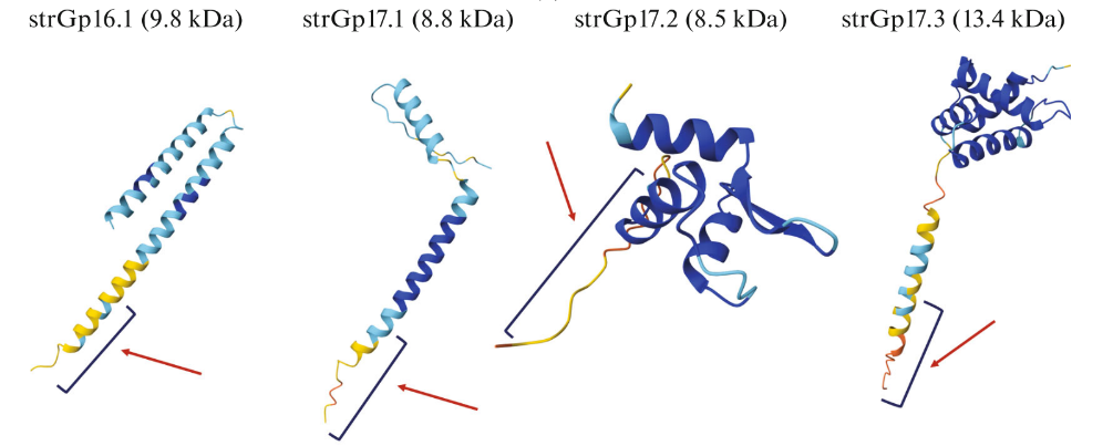
Antibiotic resistance is a major global threat, especially in the case of such gram-negative pathogens as Escherichia coli and Pseudomonas aeruginosa. Bacteriophages encode many uncharacterized proteins, making them a potential source of new antimicrobial agents. We examined early proteins of the vB_EcoP_G7C (G7C) phage for antibacterial activity. Thirteen short (<1000 bp) ORFs without known homologs were cloned into an inducible system and tested in E. coli and P. aeruginosa. Four proteins—strGp16.1, strGp17.1, strGp17.2, and strGp17.3—significantly inhibited bacterial growth, and strGp16.1 completely suppressed P. aeruginosa under experimental conditions. These findings indicate that early phage proteins can influence bacterial physiology and may provide a basis for novel antibacterial strategies. DOI
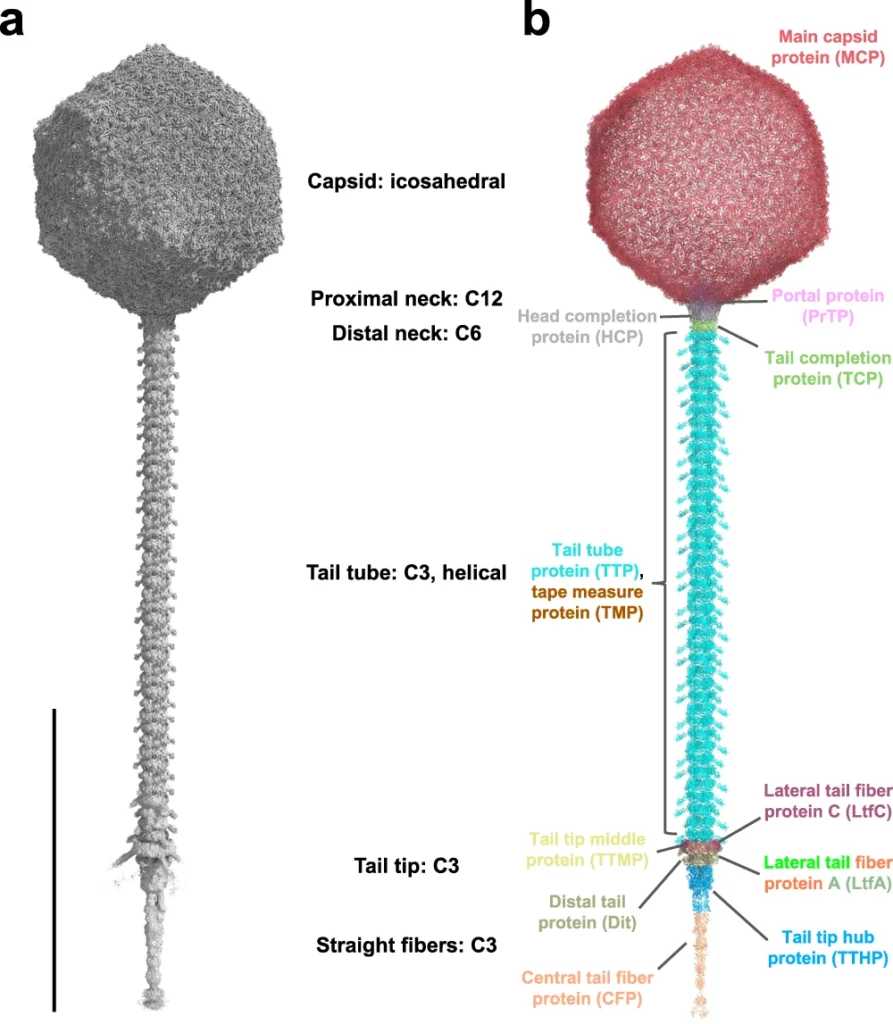
Nearly complete structure of bacteriophage DT57C reveals architecture of head-to-tail interface and lateral tail fibers.
Ayala, R., Moiseenko, A.V., Chen, T.H., Kulikov, E.E., Golomidova, A.K., Orekhov, P.S., Street, M.A., Sokolova, O.S., Letarov, A.V., Wolf, M.
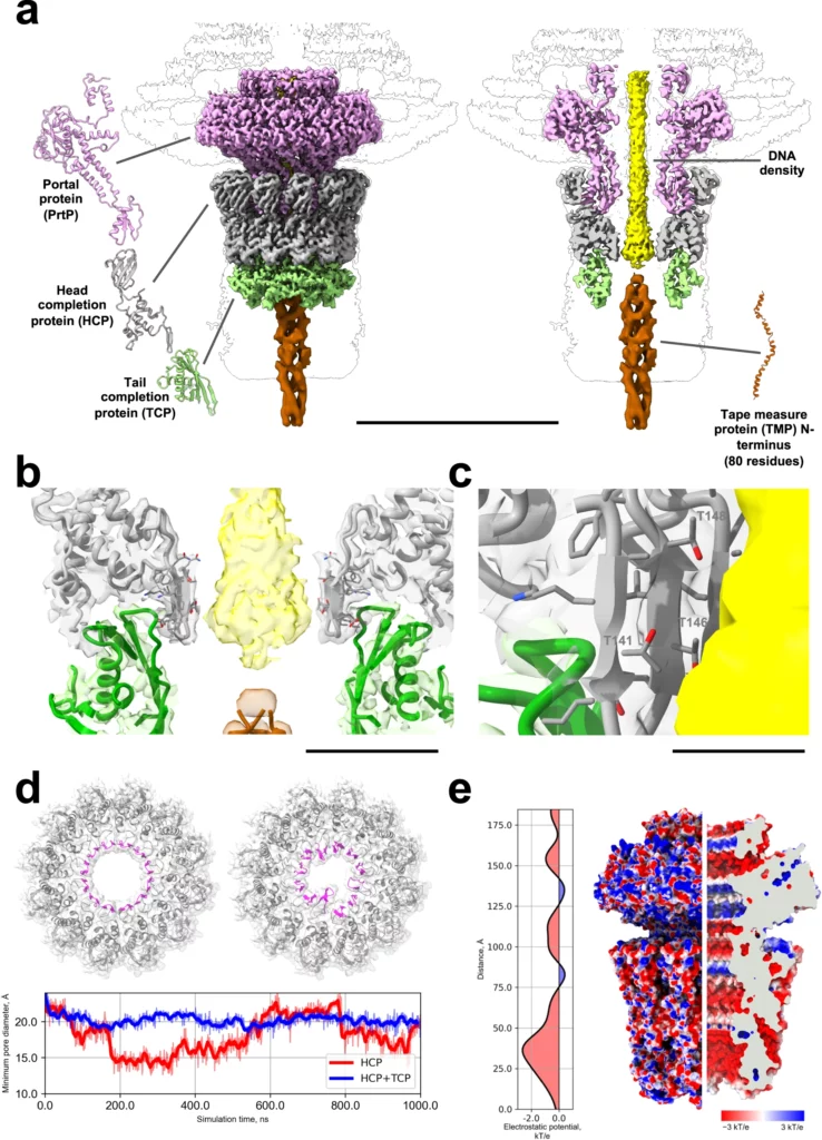
The T5 family of viruses are tailed bacteriophages characterized by a long non-contractile tail. The bacteriophage DT57C is closely related to the paradigmal T5 phage, though it recognizes a different receptor (BtuB) and features highly divergent lateral tail fibers (LTF). Considerable portions of T5-like phages remain structurally uncharacterized. Here, we present the structure of DT57C determined by cryo-EM, and an atomic model of the virus, which was further explored using all-atom molecular dynamics simulations. The structure revealed a unique way of LTF attachment assisted by a dodecameric collar protein LtfC, and an unusual composition of the phage neck constructed of three protein rings. The tape measure protein (TMP) is organized within the tail tube in a three-stranded parallel α-helical coiled coil which makes direct contact with the genomic DNA. The presence of the C-terminal fragment of the TMP that remains within the tail tip suggests that the tail tip complex returns to its original state after DNA ejection. Our results provide a complete atomic structure of a T5-like phage, provide insights into the process of DNA ejection as well as a structural basis for the design of engineered phages and future mechanistic studies. DOI
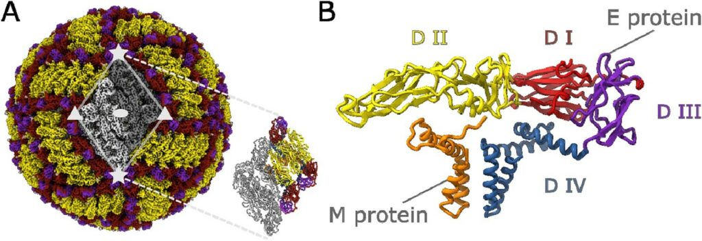
Structural diversity of tick-borne encephalitis virus particles in the inactivated vaccine based on strain Sofjin
Andrey Moiseenko, Yichen Zhang, Mikhail F. Vorovitch, Alla L. Ivanova, Zheng Liu, Dmitry I. Osolodkin, Alexey M. Egorov, Aydar A. Ishmukhametov, Olga S. Sokolova
The main approach to preventing tick-borne encephalitis (TBE) is vaccination. Formaldehyde-inactivated TBE vaccines have a proven record of safety and efficiency but have never been characterized structurally with atomic resolution. We report a cryoelectron microscopy (cryo-EM) structure of the formaldehyde-inactivated TBE virus (TBEV) of Sofjin-Chumakov strain representing the Far-Eastern subtype. A 3.8 Å resolution reconstruction reveals the structural integrity of the envelope E proteins, specifically the E protein ectodomains. The comparative study shows a high structural similarity to the previously published structures of the TBEV European subtype strains Hypr and Kuutsalo-14. A fraction of inactivated virions exhibits asymmetric features including the deformations of the membrane profile. We propose that the heterogeneity is caused by inactivation and perform a local variability analysis on the small parts of the envelope protein shell to reveal membrane curvature features possibly induced by the inactivation. The results of this study will have implications for the design of novel vaccines against diseases caused by flaviviruses. DOI